Senior Reefer yikai Posted November 24, 2010 Senior Reefer Share Posted November 24, 2010 So i just got a new microscope which allows maximum 400x magnification. Use it almost every week in school but never had the chance to use it for my own fun. so decided to take it for a little spin. i examined 3 specimens under the microscope. Bryopsis, Xenia and a clipping from my fish's tail. Quote Link to comment Share on other sites More sharing options...
Senior Reefer yikai Posted November 24, 2010 Author Senior Reefer Share Posted November 24, 2010 Here's the first subject. Bryopsis. This shot was taken under 100x magnification. I'm afraid without paraffin oil, i'm un-able to go to 400x magnification. A drop of paraffin oil is needed for 400x magnification so that the glass slide and the microscope lens is in contact with the oil, which prevents refraction of the surrounding light. anyway i don't have the oil and cooking oil won't work. so here's bryopsis under 100x magnification. The little green circles are actually chlorophyll inside the bryopsis's cell. the slide isn't properly made so the cell wall and other parts of the cell is not visible. withh 400x magnification, the individual chloroplast pigments within the chlorophyll will be visibile. unfortunately i cannot produce that. time to hunt for some paraffin oil. Quote Link to comment Share on other sites More sharing options...
Senior Reefer yikai Posted November 24, 2010 Author Senior Reefer Share Posted November 24, 2010 Here's a "petal" of xenia under 100x magnification. You can see the feather like appendages found on the arms of the xenia's polyps. each little tiny brown circle within the cells of the xenia contain zooxanthellae. without 400x magnification, this is as far as i can go. Quote Link to comment Share on other sites More sharing options...
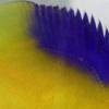
Senior Reefer yikai Posted November 24, 2010 Author Senior Reefer Share Posted November 24, 2010 And the last one is the clipping of my fish's tail. Don't worry, the fish is not harmed. a small cut is made at the outer edge of the tail and it will regenerate very quickly and the fish is not hurt. This fin clipping belongs to Cirrhilabrus marjorie, the Marjorie's fairy wrasse. You can see the bamboo like structures? Those are the rays of the fins and are covered by skin. the red spots all over are the red pigments that give the tail of the fish a reddish colour. This is the outer end of the tail which is lighter in colour. Should i cut deeper, the red colour will intensify as the number of pigments increase. and the bony rays will get thicker and bigger. this is taken under 100x magnification too. Quote Link to comment Share on other sites More sharing options...
Senior Reefer yikai Posted November 24, 2010 Author Senior Reefer Share Posted November 24, 2010 that's all for now! any requests? what should i look at next? bear in mind anything transparent like ich, flukes and other parasites need to be stained with methylene blue which i do not have. and too fine details under 400x is un-doable until i source for paraffin oil. Quote Link to comment Share on other sites More sharing options...
SRC Member AloysiusMun Posted November 24, 2010 SRC Member Share Posted November 24, 2010 Ask from your Lab technician in school. Bring a small bottle along. I am sure they wouldn't mind. Quote Link to comment Share on other sites More sharing options...
SRC Member peacemaker Posted November 24, 2010 SRC Member Share Posted November 24, 2010 Having fun with the microscope? I took a photo of a nudi once under 3x magnification. Quote Link to comment Share on other sites More sharing options...
Senior Reefer yikai Posted November 24, 2010 Author Senior Reefer Share Posted November 24, 2010 Ask from your Lab technician in school. Bring a small bottle along. I am sure they wouldn't mind. no use lah. paraffin oil is kept in a little vial with a dropper. like those iodine supplements we use for our reefs. how much can i take from her? will be better if i just bought my own. so i can use it at home. Having fun with the microscope? I took a photo of a nudi once under 3x magnification. haha. microscope is so mundane when all i look at is bacteria. this time it's fun. oh nudi! how interesting. maybe i shall look at a nudi too. or a copepod perhaps. Quote Link to comment Share on other sites More sharing options...
Eniram Posted November 24, 2010 Share Posted November 24, 2010 Wow hey lemon fantastic shots! Quote Link to comment Share on other sites More sharing options...
Eniram Posted November 24, 2010 Share Posted November 24, 2010 How about a slice of sps tissues? Quote Link to comment Share on other sites More sharing options...
Senior Reefer yikai Posted November 24, 2010 Author Senior Reefer Share Posted November 24, 2010 Wow hey lemon fantastic shots! fantastic? this consider fail leh LOL. no lah very difficult coz have to aim my camera through the tiny eye piece.... and quite blur. next time i do better. i try scale scrapings and anemone's tentacle. Quote Link to comment Share on other sites More sharing options...
Senior Reefer yikai Posted November 24, 2010 Author Senior Reefer Share Posted November 24, 2010 How about a slice of sps tissues? hmm... good idea. the best slides will be a very thin film, thin enough for all the light to pass through. but how does one acquire a thin slice of SPS tissue? Quote Link to comment Share on other sites More sharing options...
CFOh Posted November 24, 2010 Share Posted November 24, 2010 (edited) Ask from your Lab technician in school. Bring a small bottle along. I am sure they wouldn't mind. Ya, may be can bring some specimen to your school lab, take some pics under SEM.. You will see totally different world... phytoplankton Good start Edited November 24, 2010 by CFOh amend spelling Quote LFS Map in singapore __________________ ><((((º>`·.¸¸.·´¯`·.¸.·´¯`·...¸><((((º> ·´¯`·.¸. , . .·´¯`·.. >((((º> Cheers and Happy Reefing.... Link to comment Share on other sites More sharing options...
Senior Reefer yikai Posted November 24, 2010 Author Senior Reefer Share Posted November 24, 2010 Ya, may be can bring some specimen to your school lab, take some pics under SEM.. You will see totally different world... phytoplankton Good start SEM and TEM are very difficult to use. requires electron bombardment on the organism and very laychey and difficult!! i'm not trained in using SEM and TEM. most importantly, do you know how big a TEM/SEM is? it's as big as a room. from the floor to ceiling. and some are even bigger. Quote Link to comment Share on other sites More sharing options...
SRC Member AloysiusMun Posted November 24, 2010 SRC Member Share Posted November 24, 2010 SEM and TEM not easy to use. Especially the preparation of the sample. Need to use a microtome. I only used the SEM once in Singapore Polytechnic and I did not even prepare the sample. All I did was to photograph and play with the SEM. Super complex to use. Usually they have spare bottles and I don't think its expensive. Depends on how close you are to your lab tech. Hahaha. Quote Link to comment Share on other sites More sharing options...
Senior Reefer yikai Posted November 24, 2010 Author Senior Reefer Share Posted November 24, 2010 SEM and TEM not easy to use. Especially the preparation of the sample. Need to use a microtome. I only used the SEM once in Singapore Polytechnic and I did not even prepare the sample. All I did was to photograph and play with the SEM. Super complex to use. Usually they have spare bottles and I don't think its expensive. Depends on how close you are to your lab tech. Hahaha. there's many steps in sample preparation for sem and tem. need to fix, dehydrate, preserve, micro/ultratome all this. really not easy. Quote Link to comment Share on other sites More sharing options...
CFOh Posted November 24, 2010 Share Posted November 24, 2010 SEM and TEM are very difficult to use. requires electron bombardment on the organism and very laychey and difficult!! i'm not trained in using SEM and TEM. most importantly, do you know how big a TEM/SEM is? it's as big as a room. from the floor to ceiling. and some are even bigger. Ya, u should take this good opportunity to utilizes them since FOC... if not next time.. outside lab charge $3xx to $5xx at least per hour... Quote LFS Map in singapore __________________ ><((((º>`·.¸¸.·´¯`·.¸.·´¯`·...¸><((((º> ·´¯`·.¸. , . .·´¯`·.. >((((º> Cheers and Happy Reefing.... Link to comment Share on other sites More sharing options...
veliferium Posted November 24, 2010 Share Posted November 24, 2010 Ya, may be can bring some specimen to your school lab, take some pics under SEM.. You will see totally different world... phytoplankton Good start this one totally owns. Quote Link to comment Share on other sites More sharing options...
SRC Member LaW Posted November 24, 2010 SRC Member Share Posted November 24, 2010 And the last one is the clipping of my fish's tail. Don't worry, the fish is not harmed. a small cut is made at the outer edge of the tail and it will regenerate very quickly and the fish is not hurt. This fin clipping belongs to Cirrhilabrus marjorie, the Marjorie's fairy wrasse. You can see the bamboo like structures? Those are the rays of the fins and are covered by skin. the red spots all over are the red pigments that give the tail of the fish a reddish colour. This is the outer end of the tail which is lighter in colour. Should i cut deeper, the red colour will intensify as the number of pigments increase. and the bony rays will get thicker and bigger. this is taken under 100x magnification too. i like this! more please! i got 100% blue, let me know if you need. Quote If a man could beat his own fantasy. Then to only breed in captivity. Then its pointless. Genesis 1:20 And God said, Let the waters bring forth abundantly the moving creature that has life, and fowl that may fly above the earth in the open firmament of heaven. And God created great whales, and every living creature that moves, which the waters brought forth abundantly, after their kind, and every winged fowl after his kind: and God saw that it was good. And God blessed them, saying, Be fruitful, and multiply, and fill the waters in the seas, and let fowl multiply in the earth. And the evening and the morning were the fifth day. || Tank: 78" x 30" x 30" || Sump: 48" x 22" x 20" || Lights: PowerModule 10 X 80W|| Returns: 2 x HF32 || || Skimmer: BubbleKing Supermarin 300 || Wavemaker: 3 x 6100 & 1 x 6200, 2 x Wavebox 6212, WavySea || || FR: 2 x FR150 || NR: Sulphur Denitrator || CR: RM Custom Made 8" || KR: Deltec KM500 || TopUp: Tunze Osmolator 3155 || || UV: Coralife 12X 36W || Ozonizer: Sanders C200|| Controller: GHL Profilux Plus II Ex || Link to comment Share on other sites More sharing options...
SRC Member LaW Posted November 24, 2010 SRC Member Share Posted November 24, 2010 SEM and TEM not easy to use. Especially the preparation of the sample. Need to use a microtome. I only used the SEM once in Singapore Polytechnic and I did not even prepare the sample. All I did was to photograph and play with the SEM. Super complex to use. Usually they have spare bottles and I don't think its expensive. Depends on how close you are to your lab tech. Hahaha. TEM and SEM are quite easy to use. but normally its too expensive an equipment for a student to mess around with. not even in my uni. some of the samples that are published in a journal. the Main pic is the SEM, smaller one is TEM and the smallest is XRD. nice huh? all were synthesized. Quote If a man could beat his own fantasy. Then to only breed in captivity. Then its pointless. Genesis 1:20 And God said, Let the waters bring forth abundantly the moving creature that has life, and fowl that may fly above the earth in the open firmament of heaven. And God created great whales, and every living creature that moves, which the waters brought forth abundantly, after their kind, and every winged fowl after his kind: and God saw that it was good. And God blessed them, saying, Be fruitful, and multiply, and fill the waters in the seas, and let fowl multiply in the earth. And the evening and the morning were the fifth day. || Tank: 78" x 30" x 30" || Sump: 48" x 22" x 20" || Lights: PowerModule 10 X 80W|| Returns: 2 x HF32 || || Skimmer: BubbleKing Supermarin 300 || Wavemaker: 3 x 6100 & 1 x 6200, 2 x Wavebox 6212, WavySea || || FR: 2 x FR150 || NR: Sulphur Denitrator || CR: RM Custom Made 8" || KR: Deltec KM500 || TopUp: Tunze Osmolator 3155 || || UV: Coralife 12X 36W || Ozonizer: Sanders C200|| Controller: GHL Profilux Plus II Ex || Link to comment Share on other sites More sharing options...
SRC Member AloysiusMun Posted November 24, 2010 SRC Member Share Posted November 24, 2010 Cool stuff LaW! Quote Link to comment Share on other sites More sharing options...
CFOh Posted November 24, 2010 Share Posted November 24, 2010 TEM and SEM are quite easy to use. but normally its too expensive an equipment for a student to mess around with. not even in my uni. some of the samples that are published in a journal. the Main pic is the SEM, smaller one is TEM and the smallest is XRD. nice huh? all were synthesized. Agree, not difficult if compare to AFM/MRM.. Just treat it as advance high power scope after specimen sputter process done.... Not sure, I think may be different control for PHD research student.. I used to visit my colleague during her studies in NUS.. Just book the time and fill in the log book... done deal... want to further studies Quote LFS Map in singapore __________________ ><((((º>`·.¸¸.·´¯`·.¸.·´¯`·...¸><((((º> ·´¯`·.¸. , . .·´¯`·.. >((((º> Cheers and Happy Reefing.... Link to comment Share on other sites More sharing options...
Eniram Posted November 25, 2010 Share Posted November 25, 2010 hmm... good idea. the best slides will be a very thin film, thin enough for all the light to pass through. but how does one acquire a thin slice of SPS tissue? ok, why not use a scapal to slice thin cross section Quote Link to comment Share on other sites More sharing options...
SRC Member allantang Posted November 25, 2010 SRC Member Share Posted November 25, 2010 O.O wow.. more pls.. algae? like how and what do they stick themselves to glass n rock with? Quote Link to comment Share on other sites More sharing options...
CFOh Posted November 25, 2010 Share Posted November 25, 2010 Agree, not difficult if compare to AFM/MRM.. Just treat it as advance high power scope after specimen sputter process done.... Not sure, I think may be different control for PHD research student.. I used to visit my colleague during her studies in NUS.. Just book the time and fill in the log book... done deal... want to further studies The fun parts not only the pictures.. u can further analyze them via EDX to understand the components inside... Quote LFS Map in singapore __________________ ><((((º>`·.¸¸.·´¯`·.¸.·´¯`·...¸><((((º> ·´¯`·.¸. , . .·´¯`·.. >((((º> Cheers and Happy Reefing.... Link to comment Share on other sites More sharing options...


Recommended Posts
Join the conversation
You can post now and register later. If you have an account, sign in now to post with your account.